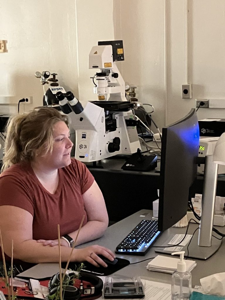
The 3i Spinning Disk Confocal Microscope is configured on a Zeiss Axio Observer Z1 automated inverted microscope platform with a motorized XY stage and piezo Z stage for rapid 3-dimensional image acquisition. The spinning disk confocal is a Yokogawa CSU-W1 T1 unit configured with a 50um pinhole disk that has increased pinhole spacing for decreased crosstalk and deep tissue imaging. Image detection is via a Prime 95B Back Illuminated Scientific CMOS camera that exhibits a 95% quantum efficiency, 11um x 11um pixel area, and a 17mm x 16mm field of view. Laser excitation includes the 445nm, 488nm, 514nm, 561nm and 637nm diode laser lines.
The Yokogawa CSU-W1 T1 confocal uses a proprietary microlens confocal disk design which provides both strong excitation of fluorophores and high-resolution confocal detection, with imaging speeds of up to 200 frames per second. The system has been optimized for rapid 2-D and 3-D imaging of live cells and tissues over time with minimal photobleaching and phototoxicity and is configured with 3i SlideBook image acquisition and analysis software and an OKO Lab environmental stage incubator for user-controlled heating and CO2.

Advanced imaging applications include:
- High-resolution, high-sensitivity confocal fluorescence imaging
- Multi-channel confocal fluorescence imaging, including standard green fluorescence (excitation 488nm), red fluorescence (excitation 561nm), and far-red fluorescence (excitation 637nm), as well as cyan fluorescence (excitation 445nm) and yellow fluorescence (excitation 514nm).
- 2D and 3D live cell imaging and quantitative analyses over time
- Multi-area and/or multi-well imaging
- Rapid 2D and 3D large area imaging
- Cell counting and image analysis software
- 3D rendering and animation
- Conventional epi-fluorescence imaging (blue, green, red)
- Transmitted light imaging (brightfield, polarized light, DIC)
- OKO Lab heating/cooling/carbon dioxide environmental stage incubator
