The Center for Advanced Microscopy (CAM) offers full comprehensive transmission electron microscopy services for the biological sciences including plant and animal samples, bacteria, and pathology specimens. In addition, full services are provided for materials science research such as polymers and plastics that require sample sectioning.
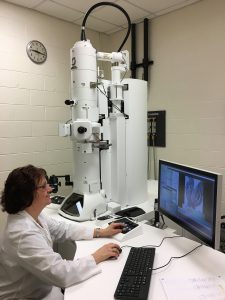
The primary transmission electron microscope for Biological Sciences TEM at the Center for Advanced Microscopy is a JEOL 1400 Flash. It has a maximum accelerating voltage of 120 kV, is equipped with a Lanthanum Hexaboride “Lab Six” emitter, and has a lattice resolution 0.2 nm using a high contrast pole piece.
The camera is a newly developed Matataki Flash sCMOS design that dramatically reduces readout noise (less than 0.9 electrons) while maintaining an extremely high frame rate of 30 frames per second. Other features include Limitless Panorama Function (automated stitching) that can cover a wide area with no limitation on the number of pixels and Optical Microscope Correlation function in which an image from an optical microscope can be imported and overlaid on a TEM image to facilitate correlative microscopy. The microscope has a four grid holder and a high tilt holder capable of plus or minus 70 degree tilts. The sample holders are interchangeable between the JEOL 1400 and the other TEM at CAM, the JEOL 2200FS.
This microscope offers excellent imaging capability for all biological samples and also for many physical science samples. In addition, the JEOL 2200FS ultra-high resolution TEM is available if needed.
The JEOL 1400 Flash TEM at the Center for Advanced Microscopy.
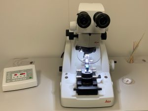
CAM has a full range of sample preparation equipment for all types of biological samples. The Leica UC7RT ultramicrotome is available for ultrathin tissue sectioning. Diamond knives for the ultramicrotome are also available for experienced users during business hours.

The Leica UC7 RT produces high quality and reproducible thin sections for biological and material sciences. Easy to use with 4 channels to store sectioning settings.

Golgi apparatus, image taken on the JEOL 1400 Flash TEM at the Center for Advanced Microscopy
CAM offers conventional TEM fixation and embedding procedures. Negative staining procedures for particles in suspension (bacteria, viruses, lipids, etc.) are available. Immunogold labeling procedures are available for specific localization of cell components. We also offer complete services for sectioning of polymers films and plastics for materials science research.
Paraffin embedding and sectioning, cryostat sectioning, and thick plastic sectioning is available to support confocal scanning laser microscopy (CLSM), laser capture microscopy (LCM), and conventional light microscopy.
We offer the following services:
- Negative staining for samples in suspension (Bacteria, virus, Lipids, etc.)
- Sample specific nanoparticle staining
- Immunogold experiments. Specific fixation and embedding procedures for each individual sample.
- Thin sectioning of thin polymer films
Additional equipment available:
- Glass knife maker (GKM, RMC, Boeckeler instruments).
For biological TEM services contact Dr. Alicia Withrow, 517-355-5004, pastorle@msu.edu.
See our Service and User charges page.
For enrollment in NSC-810 Biological TEM Lab, contact Dr. Alicia Withrow, 517-355-5004, pastorle@msu.edu. This is a limited enrollment course with specific enrollment procedures. Please see our Courses page for more information.
