Laser Capture Microscopy
The Center for Advanced Microscopy offers comprehensive Laser Capture Microscopy (LCM) services for the biological sciences including a Zeiss Palm MicroBeam IV Laser Capture Microscope and a full range of sample preparation equipment, including a Leica CM 1850 cryostat for fresh tissue sectioning, a Leica RM2125RT microtome for paraffin sectioning.
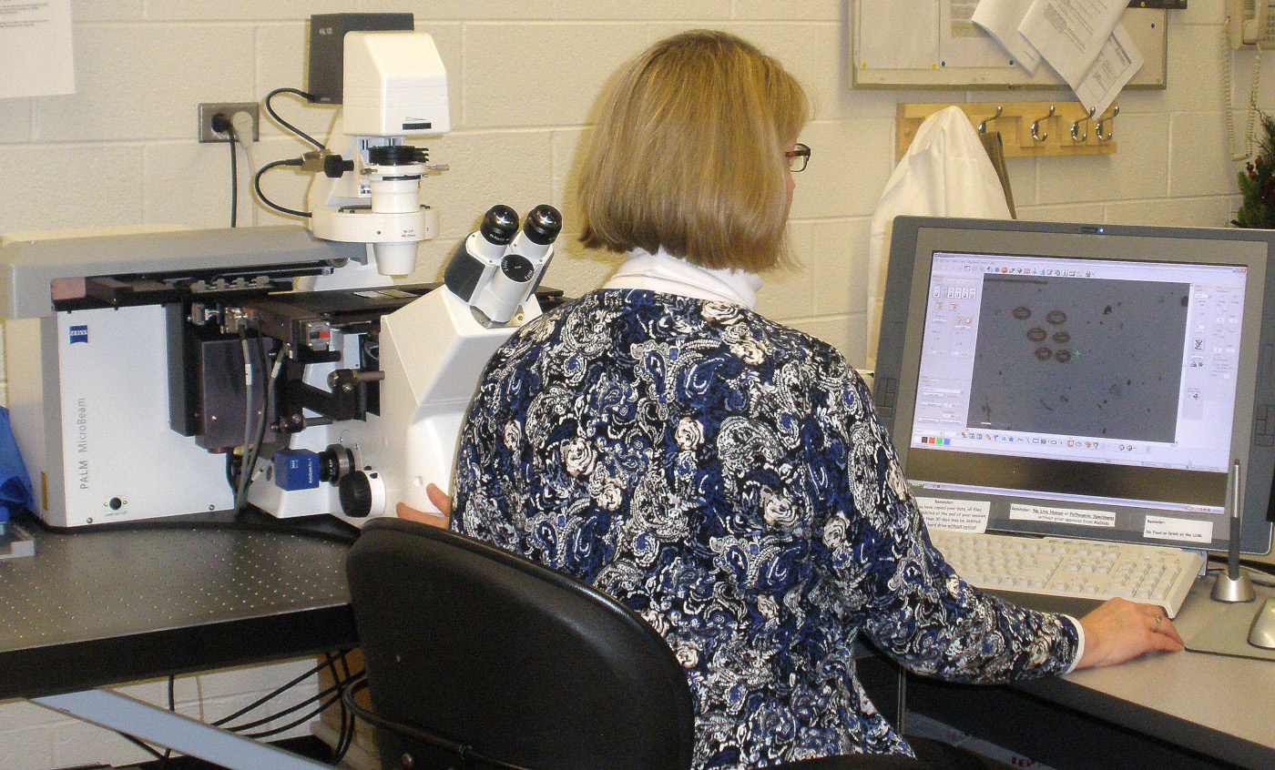
The Zeiss Palm Microbeam IV Laser Capture Microscope at the MSU Center for Advanced Microscopy
The Zeiss Palm MicroBeam IV Laser Capture Microscope provides excellent cell and tissue isolation for downstream analyses, such as RNA, DNA or protein sequencing. The LCM system is configured on a fully automated Zeiss AxioObserver Z1 inverted microscope and is equipped with a 351 nm laser for the tissue microdissection and laser capture. The system is also configured with the RoboMover automated stage, which permits synchronized movement of the sample isolation and collection tube. A full selection of objectives peremits tissue isolation at a range of magnifications in either a brightfield-imaging mode using the AxioCam ICc 1 high-resolution color camera or in a fluorescence-imaging mode using the AxioCam MRm high-resolution low-light camera.
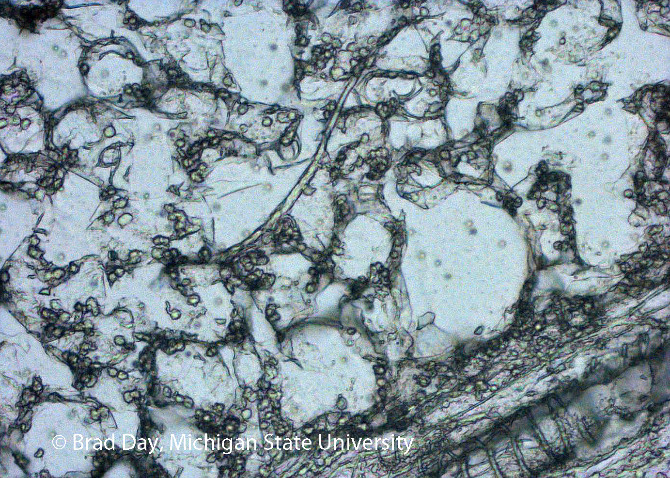
Image of sectioned leaf tissue from cucumber, Cucumis sativus, infected with downey mildew fungus, Pseudoperonospora cubensis. Image taken on the Zeiss Palm Laser Capture Microscope at the Center for Advanced Microscopy. Research from the lab of Dr. Brad Day, Michigan State University along with graduate student Alyssa Burkhardt
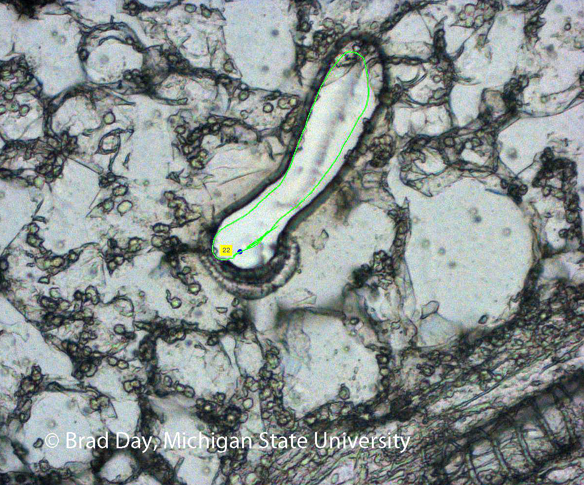
Image of the above section after cutting. The laser cut and ejected the fungal hyphal cells outlined in green, which were then sent for RNA analysis. Image taken on the Zeiss Palm Laser Capture Microscope at the Center for Advanced Microscopy. Research from the lab of Dr. Brad Day, Michigan State University along with graduate student Alyssa Burkhardt
For Laser Capture Microscope services contact Melinda Frame, framem@msu.edu, 517-432-2327. August 2025 - the Zeiss Palm microscope is currently down for repair. Please reach out if you are interested in it however!