Light Microscopy
Expand the list below for more details about each of the services offered below.
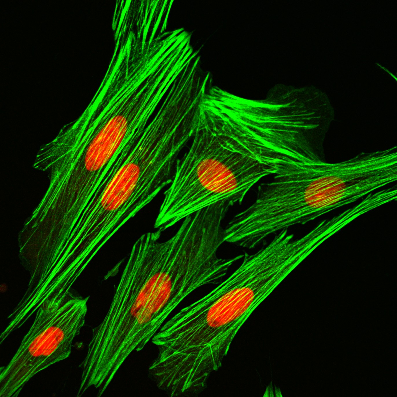
The Center for Advanced Microscopy offers a comprehensive range of advanced imaging applications, including:
- High-resolution, high-sensitivity confocal imaging in both fluorescence and reflected light modes
- Multi-channnel fluorescence imaging, including blue fluorescence (excitation 405 nm) through far red fluorescence (excitation 640 nm)
- High-speed resonance scanning
- 3D rendering and animation
- Co-localization analysis
- Cell counting and image analysis software
- Live cell imaging and quantitative analysis over time
- FRAP and FRET imaging and analysis
- Fluorescence protein imaging, including CPF, GFP, YFP, RFP, and BiFC analysis
- Spectral imaging
- High resolution large area scanning
- Total Internal Reflection Fluorescence (TIRF) microscopy
- Super Resolution STORM imaging
- Extended Depth of Focus (EDF) analysis, Topography analysis, Z height profiling
- Transmitted light imaging (brightfield, phase contrast, polarized light, DIC)
- Heated stage incubation (ambient to 99 degrees C)
- Cooling stage (ambient to -5 degrees C)
- Heating/cooling/Carbon Dioxide environmental stage incubation
For Confocal Laser Scanning Microscope services contact Melinda Frame, framem@msu.edu, 517-432-2327.
See our Service and User charges page.
Paraffin embedding and sectioning, cryostat sectioning, and thick plastic sectioning is available to support confocal scanning laser microscopy (CLSM), laser capture microscopy (LCM), and conventional light microscopy. Contact Dr. Alicia Withrow, pastorle@msu.edu, for more information.
Keyence
- Large Depth-of-Field by using Focus Stacking (Z-Stacking)
- Color Imaging from 1X to 5,000X
- Samples up to 40 mm x 40 mm can be imaged
- 3D information including profiles, roughness, and volume measurements
- Full tilt stand, up to 90 degrees
- Large area montages
For service work on the microscope, please contact Amy Albin.