The MSU Center for Advanced Microscopy has a long history at MSU, tracing back to
1956. Below are some historical details assembled by former CAM Director Dr. Stan
Flegler.
MSU SEM History
Michigan State University was one of the first universities in the country to acquire
an SEM.
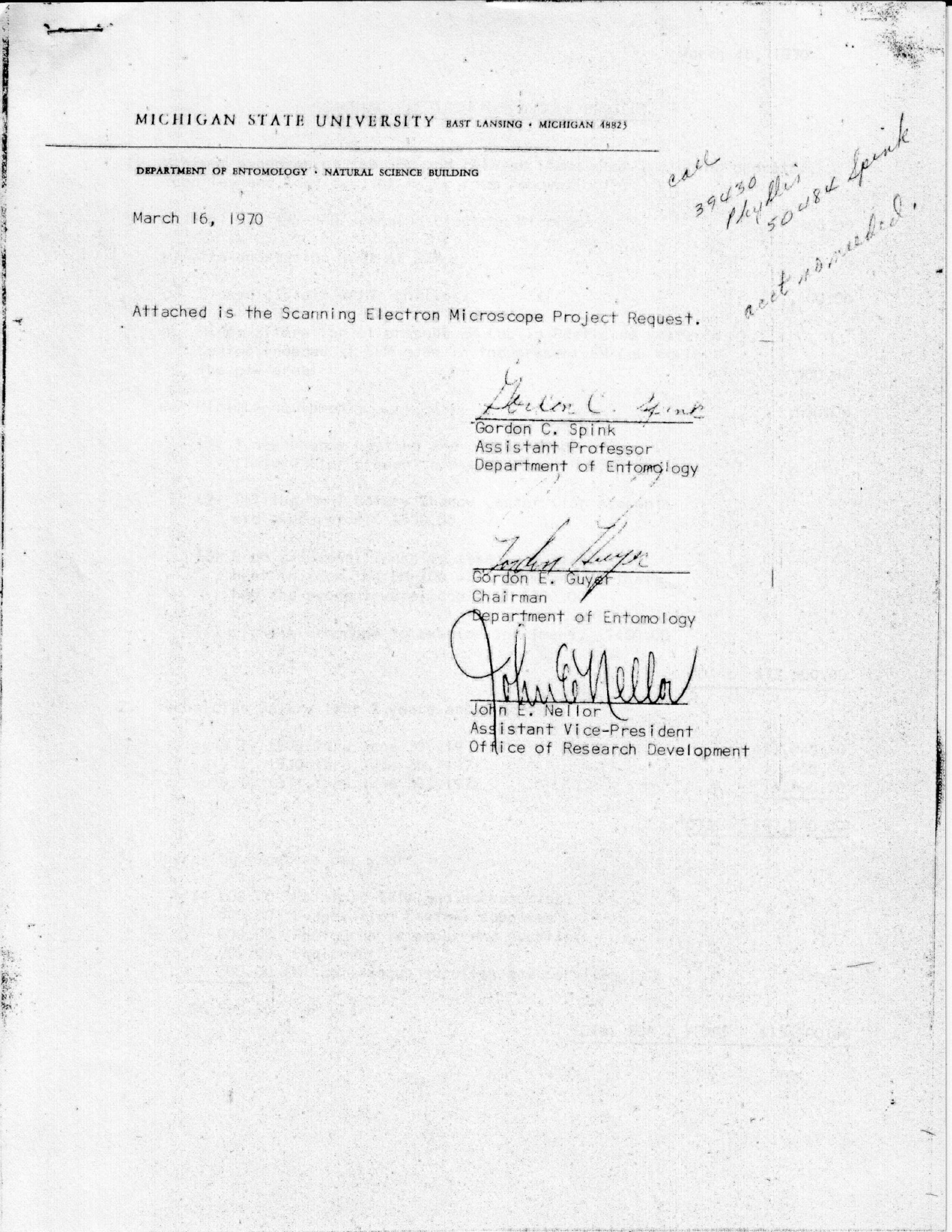
In 1970, Michigan State University had the foresight to purchase a newly developed
microscope called a scanning electron microscope or SEM. The first commercial SEM
had entered the market only five years before. The SEM that MSU purchased was the
second such microscope sold in the State of Michigan!
Fig. 1. The request for the SEM sent to Vice President of Research Dr. Milton Muelder.
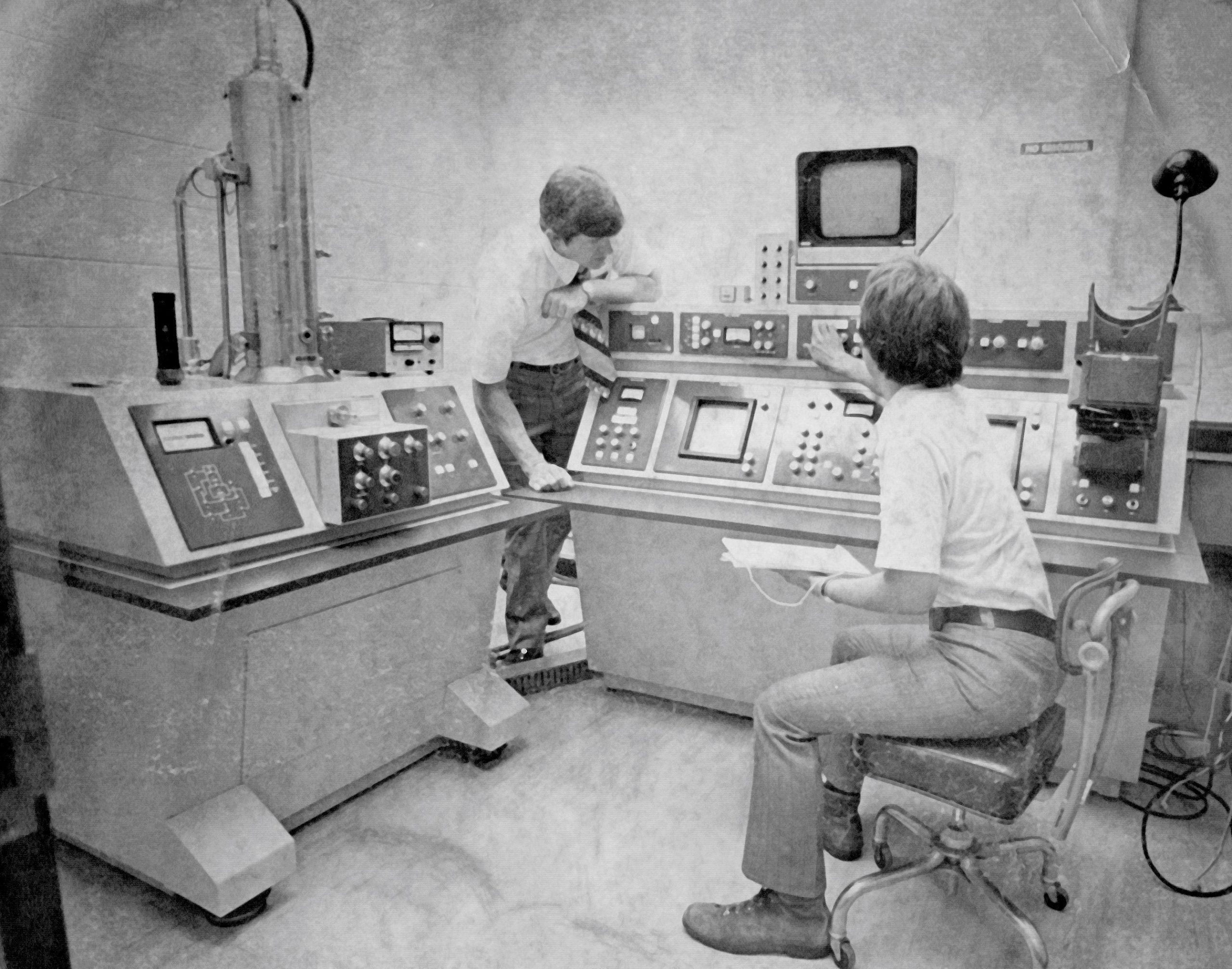
The microscope is shown in the photo below, taken in 1975. It was a model AMR-900,
produced by the Advanced Metals Research Corporation, Burlington, MA. In the photo,
the Director of CAM at the time (Dr. Gary Hooper) is standing and the SEM Supervisor
(Dr. Stanley Flegler) is seated at the right. On a good day, this microscope was capable
of images at 5,000x. The current high resolution JEOL 7500F SEM at CAM can achieve
images at 1,000,000x.
Fig. 2. The AMR-900 SEM.
Formal classroom instruction on the microscope by Director Dr. Gary Hooper began in
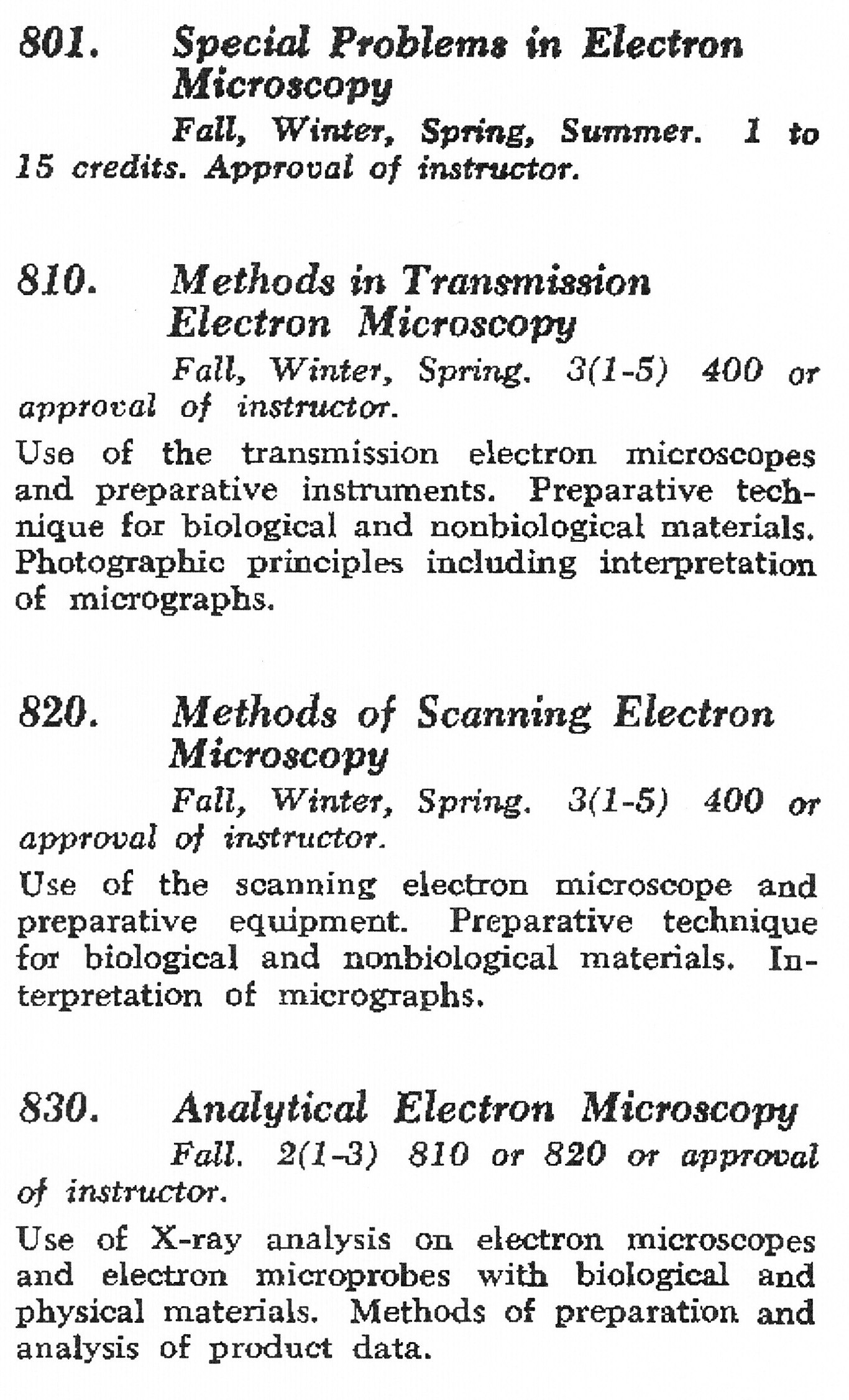
1973 under NSC-801 Special Problems in Electron Microscopy. In 1976, the class NSC-820
Methods of Scanning Electron Microscopy was established. In 1980 instruction in the
class was passed to the new Director Dr. Karen Klomparens. In 1981 instruction in
NSC-820 was passed to the SEM Supervisor Dr. Stanley Flegler who has been instructor
of record for 43 years. Over the years, over 1,400 graduate students have received
formal SEM instruction at CAM with an emphasis on underlying theory and individual
one-on-one instruction.
The 1976 Descriptions of Courses showing the newly added NSC-820 class.
MSU TEM and CAM History
Michigan State University was one of the first universities in the country to purchase
a transmission electron microscope (TEM) and make it available as a campus core facility.
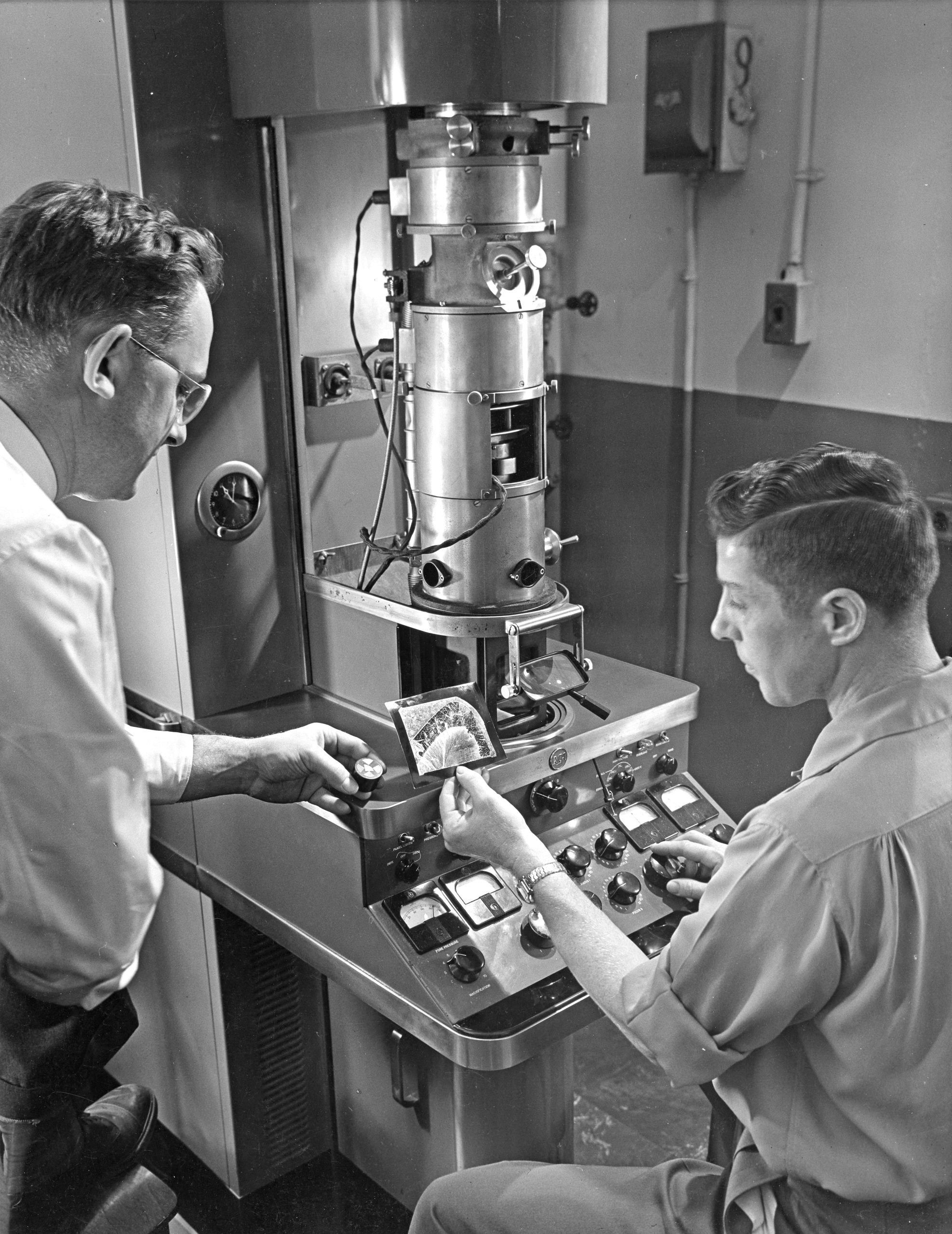
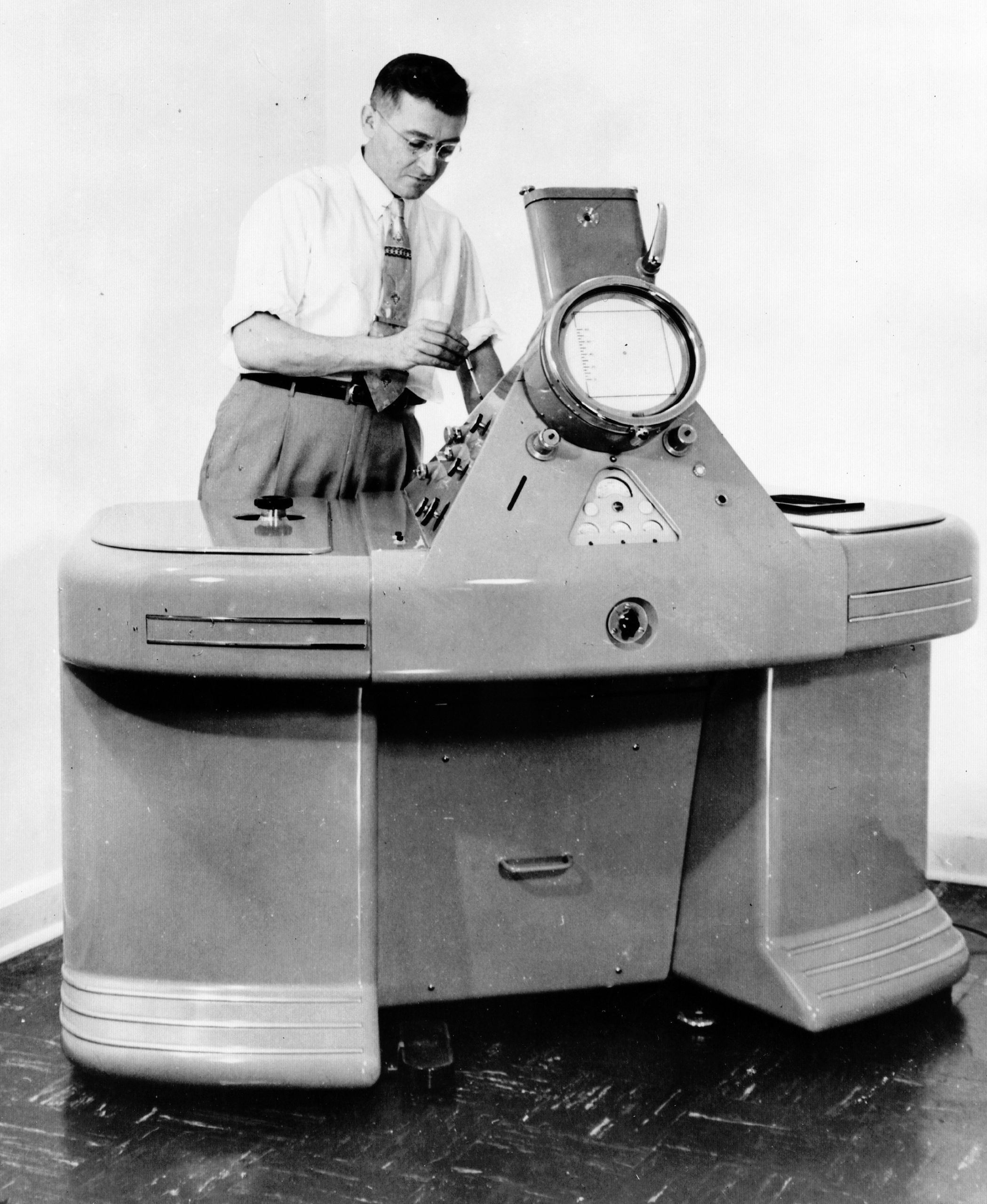
It began in 1950 when several researchers in the College of Natural Science secured
funding to acquire a transmission electron microscope (TEM). They selected the model
EMU-2, built by the Radio Corporation of America (RCA). The EMU-2 was a well established
model with an excellent reputation. The microscope was located in the basement of
the West wing of the Natural Science Building.
An RCA model EMU-2 TEM.
As time permitted, the researchers began assisting others on campus with requests
to look at their samples. In 1956, a more formal basis was established and the lab
became known as theElectron Optics Lab.
This continued until 1960, when MSU recognized that access to a newer TEM with full
time operator assistance had become vital to the campus research effort. A Philips
model EM-100 was purchased and located in the basement of the newly constructed Biology
Research Center.
A Philips EM-100 TEM.
Dozens of students were taught on the EM-100 microscope in the time frame of 1960
to 1976, when the EM-100 was decommissioned
A Director was appointed in 1960, Dr. Lee Vern Leak, and a full time technician was
hired, Ms. June Mack. Dr. Leek left MSU in 1962. He went on to do pioneering work
in the ultrastructure of the lymphatic system and would become the chair of the Anatomy
Department at Howard University. Other directors have been:
Dr. Gordon Spink 1962 to 1970.
Dr. H. Paul Rasmussen, 1970 to 1972.
Dr. Gary Hooper, 1972 to 1980.
Dr. Karen Klomparens, 1980 to 1998.
Dr. Stanley Flegler, 1998 to 2024.
The first director, Dr. Lee Vern Leak.
During the 1960’s, many MSU researchers were introduced to the capabilities of TEM
and used the Philips EM-100 at the Biology Research Center. During that time period,
major gains in microscope design and resolution were occurring. In 1968, MSU made
a decision to construct a well designed electron microscope lab in the basement of
the newly constructed Pesticide Research Center (now the Center for Integrated Plant
Systems building). A Philips EM-300 TEM was purchased for the new lab, along with
the Philips EM-100, relocated from the Biology Research Center. A scanning electron
microscope (SEM) was also located in the building. Please seeseparate articlefor information on this microscope.
The Philips EM-300 with Director Dr. Gary Hooper at the controls.
A new Director, Dr. Gary Hooper was hired in 1972. In 1973, along with input from
the electron microscope advisory committee, the name of the all-campus facility was
changed toThe Center for Electron Optics(CEO).
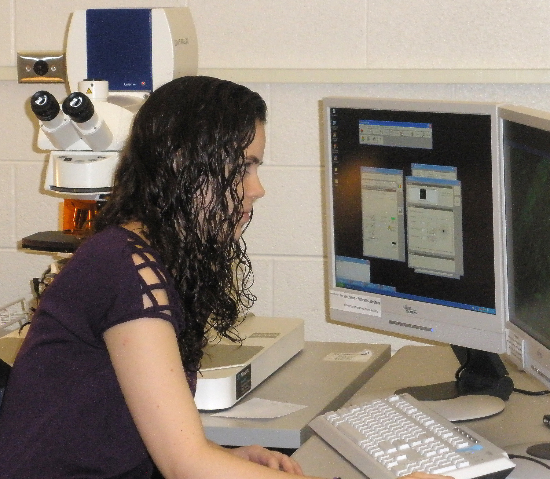
During the early 1990’s, major advances were made in the development of confocal laser
scanning microscopes (CLSM). Dr. Joanne Whallon, Department of Crop and Soil Sciences,
a major user of the CEO, secured funding for the purchase of a Zeiss LSM-210 and located
it in the Plant and Soil Sciences Building. The lab was named the Laser Scanning Microscopy
Laboratory with Dr. Whallon as Director. The lab was merged physically and administratively
with the CEO on Jan. 1, 2000. The combined lab was renamed theCenter for Advanced Microscopy.
A Zeiss LSM-210 confocal laser scanning microscope. Photo courtesy of eTech Surplus.
The user base at CAM has grown rapidly since 2000. The growth has advanced in all
four core areas, SEM, physical sciences TEM, biological sciences TEM, and CLSM (confocal
laser scanning microscopy). CAM serves the entire campus with users from nine colleges.
CAM was named aUniversity Core Facilityin 2000 by Robert Huggett, Vice President for Research and Graduate Studies. The most
recent data for calendar year 2023 showed 446 research users from 39 units on campus.
Number of unique CAM users between 2000 and 2022.
The Center for Advanced Microscopy has been serving the microscopy needs of MSU researchers
since 1956. Many of the significant research discoveries made at the University over
the years have relied on the services and microscopes at the Center.